
introduction to cardiovascular sonography
Cardiovascular sonography is a non-invasive imaging method that allows doctors to evaluate the health of the heart and blood vessels using ultrasonic technology. It is a critical tool for evaluating heart conditions early as cardiovascular disease can lead to significant morbidity and mortality.
What Is Cardiovascular
Cardiovascular sonography, also called echocardiography or vascular ultrasound, uses high-frequency sound waves to produce images of the heart and blood vessels. It allows the clinician to measure blood flow, assess blockages, and evaluate heart function without needing surgery or radiation. According to the American Heart Association, regular heart imaging helps detect cardiovascular issues early and supports better long-term outcomes.
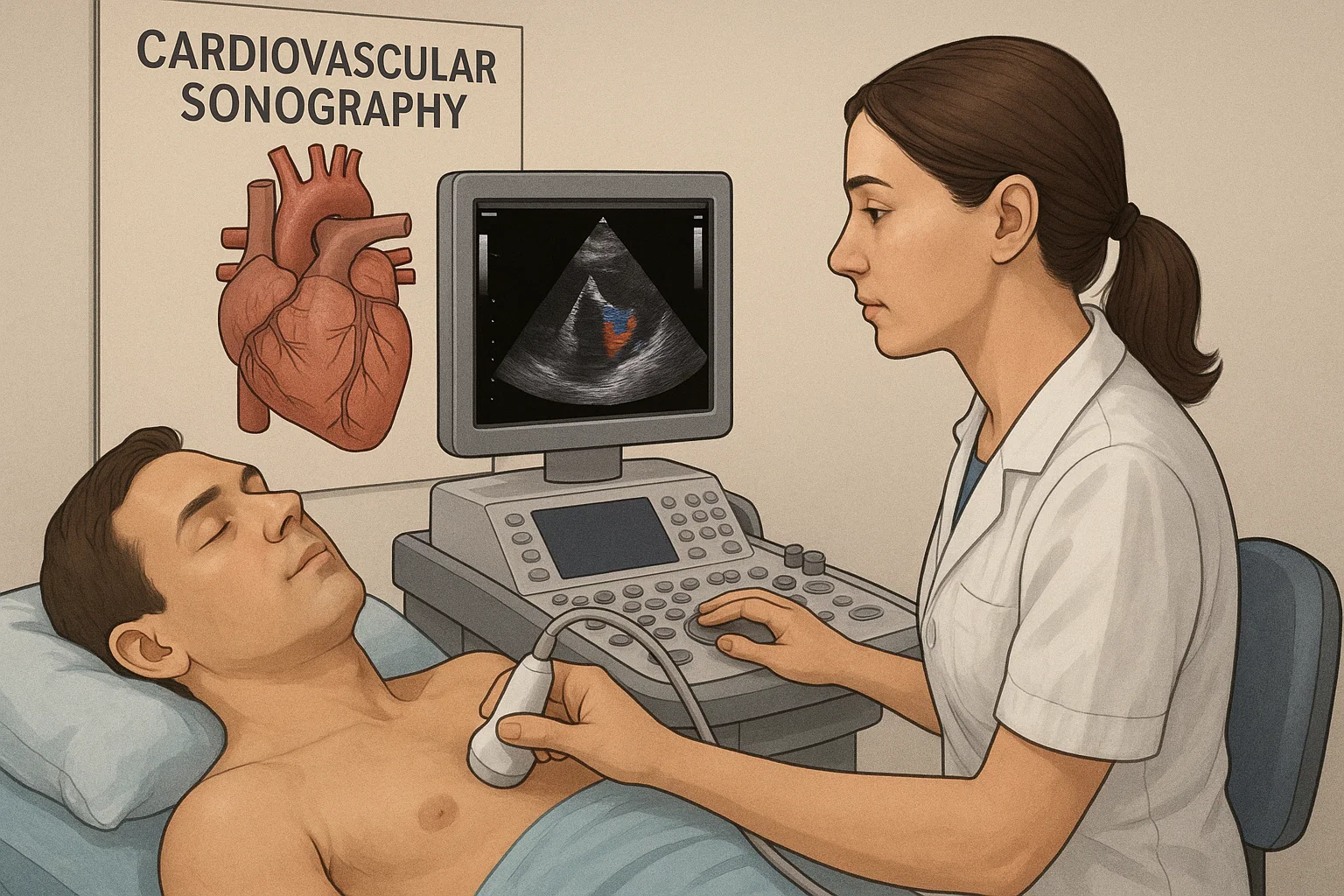
How It Works
A sonographer will place a gel on the patient’s chest or body part.
A transducer/transducer sends sound waves into the body.
The sound waves return and create images on a monitor.
This is a safe, short, and painless procedure.
Advantages of Cardiovascular Sonography
Identifies heart defects and blockages at an early stage
Aids in treatment decisions for heart failure, stroke, and more
Provides updates on recovery after heart surgery
Helps quantifying blood pressure in veins and arteries
Cardiovascular Sonography vs. Other Imaging Tests
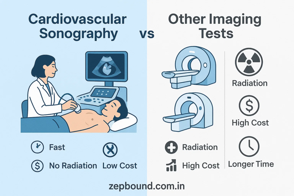
Cardiac MRI: Provides detailed 3D images but at a higher cost and is less commonly available.
CT Angiography: Nice to see blood vessels but exposes the patient to radiation.
Electrocardiogram (ECG): Measures the electrical activity of the heart but does not provide actual images of the heart structures.
Cardiovascular sonography offers major advantages: it is fast, relatively inexpensive, and uses no radiation. For more details, you can see this source: https://zepbound.com.in/cardiovascular-sonography/
Forms of Cardiovascular Sonography
Transthoracic Echocardiography (TTE)
The most frequently used echocardiography. A probe is placed on the chest to get images through the chest wall
Transesophageal Echocardiography (TEE)
A specialized test that enters a thin probe into the esophagus to obtain closer and clearer images of the heart
Stress Echocardiography
Performed before and after exercise to measure how the heart responds to physical activity
Vascular Sonography
Focused on arteries and veins, checking for blood clots, narrowing, or other vascular diseases.
Lessens the need for invasive assessments
The American Heart Association states that an early diagnosis developed through an ultrasound can increase treatment options by up to 40%.
Cardiovascular Sonography Applications
Evaluating valvular function and heart chambers
Checking for blood clots or an aneurysm
Measuring blood flow speed and direction
Evaluating potential effects of high blood pressure
Evaluating patients with pacemakers or implantable devices
Cardiovascular Sonography applications can be found in hospitals, clinics, and cardiovascular research centers.
Risks of Cardiovascular Sonography
Cardiovascular sonography is safe and does not involve radiation. While some risk is present, it is minimal.
Some patients may experience mild discomfort from the pressure of the transducer.
Also, transesophageal echocardiography, a specific procedure of echocardiography, may cause some throat irritation.
No adverse long-term effects have been established.
Comparison With Other Imaging Methods
| Imaging Type | Uses Radiation | Invasive | Common Use |
|---|---|---|---|
| Cardiovascular Sonography | No | No | Routine heart checks |
| CT Angiography | Yes | No | Detailed artery scans |
| Cardiac MRI | No | No | Advanced tissue imaging |
| Cardiac Catheterization | Yes | Yes | Diagnostic and treatment use |
Helpful Patient Tips
Dress in comfy clothes for your exam.
Refrain from consuming a heavy meal for a transesophageal procedure.
Follow any preparation instructions from your doctor.
Explain to the technician any history of heart sensation and medications you are taking.
Summary / Takeaway
Cardiovascular sonography is an ultrasound imaging technique used to evaluate the heart and blood vessels.
It is safe, noninvasive, and uses no radiation.
Cardiovascular sonogram is an ideal study to evaluate early signs of heart disease.
Cardiovascular sonograms are an important tool for treatment planning and monitoring recovery.
Cardiovascular sonograms result in real-time diagnostic images.
FAQs
- Is cardiovascular sonography painful?
No. The procedure is comfortable and painless. - How long does a sonography test take?
Most tests take 30 to 60 minutes depending on the complexity of the case. - Can it detect all problems of the heart?
It detects most structural problems and those concerning blood flow. Sometimes, a physician will recommend additional studies to complete the evaluation. - Is cardiovascular sonography safe during pregnancy?
Yes. Sound waves are used, not radiation, making it safe for patients of all ages. - Who does the test?
A certified cardiovascular sonographer, or ultrasound technician, performs the test under the supervision of a cardiologist. - How often should I receive a sonogram of my heart?
If you have been diagnosed with heart disease or have other risk factors, your physician may recommend every 6 to 12 months.