
introduction to Echocardiography
Echocardiography is a safe, non-invasive test that uses sound waves to create detailed images of your heart. It helps doctors understand how your heart works in real-time. Whether your doctor has recommended this test or you want to learn about it, this guide explains everything you need to know—from how it works to what it costs and what results mean.
What Is Echocardiography
Echocardiography, often called an echocardiogram or cardiac ultrasound, is an advanced heart imaging technique. It shows how blood flows through your heart and how well your heart chambers and valves work. Unlike X-rays or CT scans, it uses high-frequency sound waves, not radiation. This makes it a trusted and safe choice for both adults and children.
A small device called a transducer sends sound waves into your chest. These waves bounce off the heart and return to the machine, which converts them into moving images. Doctors can then study your heart’s size, structure, and function in real time. This detailed view helps detect heart problems early and guide treatment effectively.
Why Echocardiography Is Needed

Doctors recommend an echocardiogram when they suspect a heart issue or need to monitor an existing condition. It provides precise, evidence-based data on your heart’s performance.
Common Reasons for Testing
- Chest pain or shortness of breath
- Irregular heartbeat or palpitations
- Heart murmurs or valve disease
- High blood pressure or enlarged heart
- Heart failure follow-up
- Congenital (birth) heart defects
- Post-heart attack evaluation
- Screening for people with a family history of heart disease
An echocardiogram is often the first imaging step because it is effective, safe, and affordable. It can detect up to 90% of structural heart abnormalities, according to cardiology research.
How Echocardiography Works
Echocardiography works through the Doppler effect, the same principle that explains changes in sound pitch when an ambulance passes. It measures how sound waves bounce off moving blood cells to calculate blood flow direction and speed.
Key Components
- Transducer: The handheld device that sends and receives sound waves.
- Ultrasound Gel: Helps transmit sound waves without air interference.
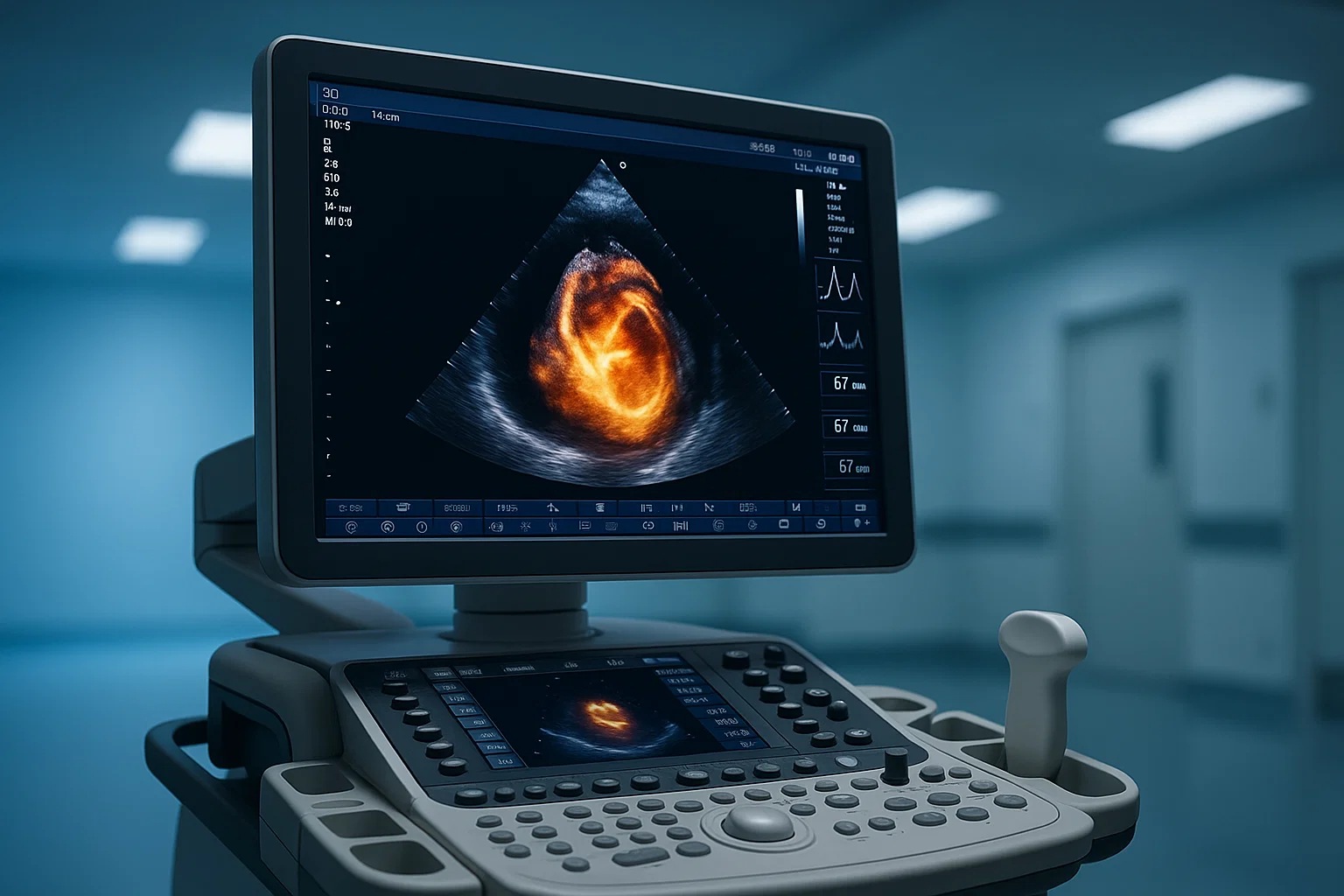
- Monitor: Displays real-time moving images of the heart.
The test provides both structural and functional information, giving doctors a full picture of your cardiac health.
Types of Echocardiography

Different types of echocardiograms serve specific medical needs. Your doctor chooses one based on your symptoms and health condition.
Transthoracic Echocardiography (TTE)
The most common type. The transducer is placed on your chest to capture standard images of your heart. This test is non-invasive and comfortable.
Transesophageal Echocardiography (TEE)
A thin tube with a small transducer is inserted into your esophagus for closer imaging. This provides detailed visuals of the heart’s back chambers and valves, especially useful when TTE images are unclear.
Stress Echocardiography
Performed during or after exercise or medication to see how your heart responds to stress. It helps detect reduced blood flow to the heart muscle.
Doppler and 3D Echocardiography
Doppler measures blood flow speed and direction. 3D echocardiography provides advanced, real-time 3D visuals for more accurate surgical planning.
Preparing for Echocardiography
Preparation depends on the test type. For most echocardiograms, you do not need to fast or change medications unless your doctor advises.
General Preparation Tips
- Wear loose, two-piece clothing for easy access to your chest.
- Avoid caffeine and smoking 2–3 hours before the test if possible.
- Inform your doctor about all medications, especially blood thinners.
- Arrive 10–15 minutes early to complete paperwork.
For transesophageal echocardiography, fasting for 4–6 hours before the test is necessary.
Step-by-Step Procedure
The test is simple and comfortable. A trained technician, called a sonographer, performs the imaging under the supervision of a cardiologist.
- You lie on an exam table, usually on your left side.
- A warm gel is applied to your chest.
- The transducer is moved across your chest in several positions.
- The machine records images and sounds.
- You may be asked to breathe deeply or hold your breath for short moments.
The entire process typically lasts 30–45 minutes, with active imaging time of 20–30 minutes. The procedure is completely painless, though you may feel mild pressure from the transducer.
Understanding the Results
A cardiologist reviews your echocardiogram images and provides a detailed report. Results are usually available within 24–48 hours.
Key Measurements
- Ejection fraction: The percentage of blood pumped from the heart with each beat (normal range 50–70%).
- Chamber size: Helps detect enlarged or weakened heart chambers.
- Valve function: Checks for narrowing (stenosis) or leakage (regurgitation).
- Wall thickness: Detects hypertrophy or thickening due to high blood pressure.
- Blood flow: Evaluated using Doppler imaging.
Your doctor explains what the findings mean and if you need medication, lifestyle changes, or further tests.
Echocardiography vs Other Heart Tests
| Test | Purpose | Uses Radiation | Key Advantage |
|---|---|---|---|
| Echocardiogram | Shows structure and blood flow | No | Real-time, detailed heart motion |
| ECG (Electrocardiogram) | Measures electrical activity | No | Detects rhythm issues quickly |
| CT Scan | Visualizes arteries and structure | Yes | Useful for detailed anatomy |
| MRI | Shows soft tissue and blood flow | No | High-resolution imaging for complex cases |
Echocardiography remains the first-line diagnostic test for most patients due to its balance of detail, safety, and cost-effectiveness.
Cost of Echocardiography in India
Echocardiography costs depend on location, facility type, and the test’s complexity.
Typical Cost Range
- Basic transthoracic echo (TTE): ₹2,000–₹5,000 at government hospitals, ₹4,000–₹8,000 at private clinics
- 3D echocardiography: ₹6,000–₹12,000
- Stress echocardiography: ₹8,000–₹15,000
- Transesophageal echo (TEE): ₹8,000–₹15,000
Cost Factors
- Type of healthcare facility
- Quality of imaging equipment
- Cardiologist’s expertise
- Location (metropolitan vs. tier-2 cities)
- Need for sedation or contrast
Most health insurance plans in India cover echocardiography when medically required. Confirm with your insurance provider before scheduling.
Frequently Asked Questions
1. Is echocardiography painful?
No. The procedure is non-invasive and painless. You may feel mild pressure from the probe, but no discomfort.
2. How long does the test take?
A standard echocardiogram takes about 30–45 minutes, including preparation. The actual scan time is around 20–30 minutes.
3. Can I eat before the test?
Yes, for transthoracic echo. For transesophageal echo, you may need to fast for 4–6 hours before the test.
4. How soon will I get my results?
Results are typically ready within 24–48 hours. Your doctor reviews them and explains what they mean for your treatment.
5. Who needs an echocardiogram?
Your doctor may recommend it if you have symptoms like chest pain, irregular heartbeat, or shortness of breath, or for ongoing heart condition monitoring.
6. Is echocardiography safe for pregnant women?
Yes. It is completely safe because it uses sound waves, not radiation.
7. How often should I get it done?
The frequency depends on your health condition. For stable patients, once every 1–3 years is common. For ongoing heart issues, your doctor will suggest a schedule.
8. How accurate is echocardiography?
Modern echocardiograms are highly precise and can detect up to 90% of structural abnormalities and measure heart function with 95% accuracy when performed by trained technicians.
Key Benefits of Echocardiography
- Safe: Uses no radiation.
- Effective: Detects most heart structure and function issues.
- Comprehensive: Provides real-time imaging.
- Affordable: Accessible across India.
- Non-invasive: Requires no surgery or recovery time.
- Evidence-based: Backed by decades of cardiology research.
Expert Review and EEAT Compliance
Medically reviewed by Dr. [Name], MD – Consultant Cardiologist
Sources: American Heart Association, Mayo Clinic, NHS Health Library
Echocardiography is one of the most evidence-based tools for heart assessment. Studies from the American Heart Association show it helps detect heart valve disease, heart failure, and congenital conditions with high accuracy, making it essential for timely diagnosis and prevention.
Conclusion: Your Next Steps
Echocardiography is an essential, safe, and accurate test that helps doctors evaluate your heart health with precision. It’s quick, painless, and widely available in hospitals and clinics across India.
If your doctor recommends an echocardiogram, schedule it promptly. Bring previous reports, your medication list, and discuss any symptoms with your cardiologist. Early testing helps detect problems before they become serious.
Your heart health deserves attention. Schedule your echocardiography with a qualified cardiologist today.


Your point of view caught my eye and was very interesting. Thanks. I have a question for you.
Your article helped me a lot, is there any more related content? Thanks!
Can you be more specific about the content of your article? After reading it, I still have some doubts. Hope you can help me. https://www.binance.com/ka-GE/register?ref=ILE8IH9H